|
Anatomy of
thoracic CT Unlike a chest
X-ray, one should be able to account for everything
that is seen on a thoracic CT scan as one is
looking at an almost 2-dimensional structure (i.e.
slice) rather than a composite of a 3-dimensional
object squashed into 2 dimensions. The basic
composition of a structure can be ascertained by
the level of greyness, e.g. bone tends to be white,
air black. Remember however that windowing can
affect markedly the level of grey of a particular
tissue. If in doubt, the easiest solution is to
compare the level of grey of unknown tissue with
that of known tissue. Deciding on exactly
which structure one is looking at depends on
knowledge of anatomy. Much of this is quite basic
but the mediastinum is complex and requires more
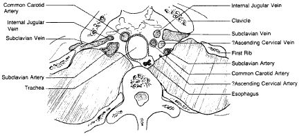
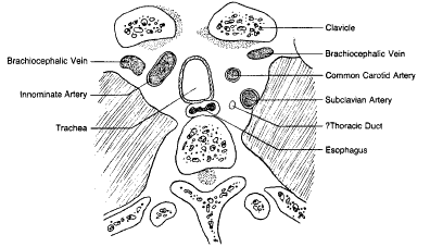
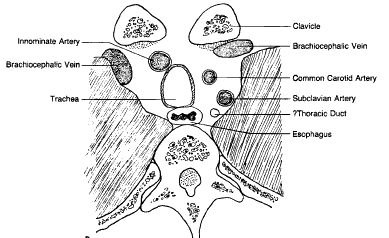
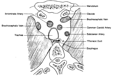
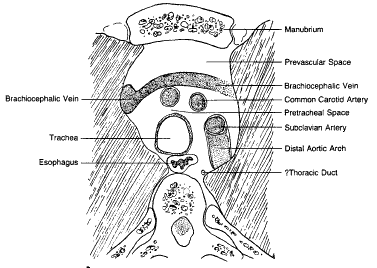
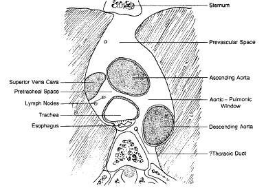
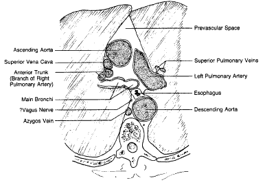
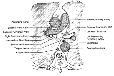
detailed knowledge. The following line drawings are
tracings of actual CT scans and depict the
arrangement of the mediastinal structures at
different levels on axial imaging. Labels preceded
by a question mark reflect the limitation of
spatial resolution on the scans
analysed.
All concepts, information and graphics contained within this internet site are the sole copyright(©) of M,A&A (©M,A&A)
|
