|
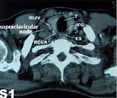
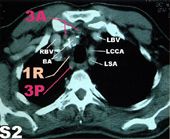
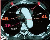
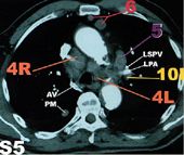
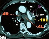
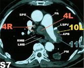
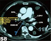
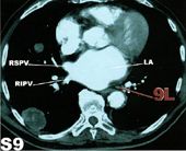
To put this anatomy
into practice there follows a series of scans of a
patient with diffuse metastatic rectal carcinoma
involving all the major lymph node groups. Contrast
has been given to give maximum delineation of the
vessels. These examples have been taken from a
chart published in Radiographics 1999, Volume 19,
which demonstrates the American Joint Committee on
Cancer (AJCC) and Union Internationale Contre le
Cancer (UICC) regional lymph node classification
for lung cancer staging. A key to the abbreviations
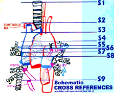
is appended below. The following diagram shows
the level of the CT sections:
All concepts, information and graphics contained within this internet site are the sole copyright(©) of M,A&A (©M,A&A)
|

